| |
| |
Caption: Book - Research on Campus (1949)
This is a reduced-resolution page image for fast online browsing.
EXTRACTED TEXT FROM PAGE:
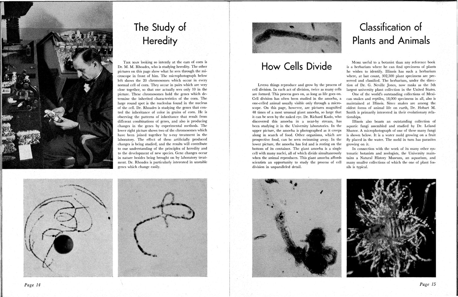
The Study of Heredity T H E MAN looking so intently at the ears of corn is Dr. M. M. Rhoades, who is studying heredity. T h e other pictures on this page show what he sees through the microscope in front of him. T h e microphotograph below left shows the 20 chromosomes which occur in every normal cell of corn. They occur in pairs which are very close together, so that one actually sees only 10 in the picture. These chromosomes hold the genes which determine the inherited characteristics of the corn. T h e large round spot is the nucleolus found in the nucleus of the cell. Dr. Rhoades is studying the genes that control the inheritance of color in grains of corn. He is observing the patterns of inheritance that result from different combinations of genes, and also is producing changes in the genes by experimental methods. T h e lower right picture shows two of the chromosomes which have been joined together by x-ray treatment in the laboratory. T h e effect of these artificially produced changes is being studied, and the results will contribute to our understanding of the principles of heredity and to the development of new species. Gene changes occur in nature besides being brought on by laboratory treatment. Dr. Rhoades is particularly interested in unstable genes which change easily. Classification of Plants and Animals How Cells Divide LIVING things reproduce and grow by the process of cell division. In each act of division, twice as many cells are formed. This process goes on, as long as life goes on. Cell division has often been studied in the amoeba, a one-celled animal usually visible only through a microscope. O n this page, however, are pictures magnified 48 times of a most unusual giant amoeba, so large that it can be seen by the naked eye. Dr. Richard Kudo, who discovered this amoeba in a near-by stream, has been studying it in the University laboratories. I n the upper picture, the amoeba is photographed as it creeps along in search of food. Other organisms, which are prospective food, can be seen swimming away. In the lower picture, the amoeba has fed and is resting on the bottom of its container. T h e giant amoeba is a single cell with many nuclei, all of which divide simultaneously when the animal reproduces. This giant amoeba affords scientists an opportunity to study the process of cell division in unparalleled detail. M O R E useful to a botanist than any reference book is a herbarium where he can find specimens of plants he wishes to identify. Illinois has such a herbarium where, at last count, 302,500 plant specimens are preserved and classified. T h e herbarium, under the direction of Dr. G. Neville Jones, now ranks as the 5th largest university plant collection in the United States. One of the world's outstanding collections of Mexican snakes and reptiles, 18,000 specimens in all, also is maintained at Illinois. Since snakes are among the oldest forms of animal life on earth, Dr. Hobart M. Smith is primarily interested in their evolutionary relationships. Illinois also boasts an outstanding collection of aquatic fungi assembled and studied by Dr. Leland Shanor. A microphotograph of one of these many fungi is shown below. It is a water mold growing on a fruit fly placed in the water. T h e mold in turn has parasites growing on it. In connection with the work of its many other systematic botanists and zoologists, the University maintains a Natural History Museum, an aquarium, and many smaller collections of which the one of plant fossils is typical. Page 15
| |