| |
| |
Caption: Book - Research on Campus (1949)
This is a reduced-resolution page image for fast online browsing.
EXTRACTED TEXT FROM PAGE:
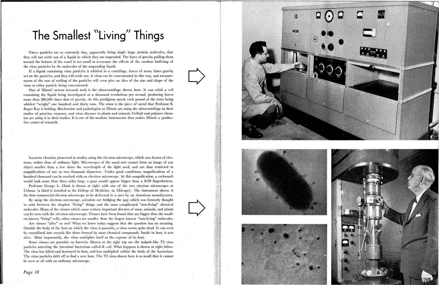
The Smallest Living Things V I R U S particles are so extremely tiny, apparently being single large protein molecules, that they will not settle out of a liquid in which they are suspended. T h e force of gravity pulling them toward the bottom of the vessel is too small to overcome the effects of the random buffeting of the virus particles by the molecules of the suspending liquid. If a liquid containing virus particles is whirled in a centrifuge, forces of many times gravity act on the particles, and they will settle out. A virus can be concentrated in this way, and measurements of the rate of settling of the particles will even give an idea of the size and shape of the virus or other particle being concentrated. One of Illinois' newest research tools is the ultracentrifuge shown here. It can whirl a cell containing the liquid being investigated at a thousand revolutions per second, producing forces more than 260,000 times that of gravity. At this prodigious speed, each pound of the rotor being whirled "weighs" one hundred and thirty tons. T h e rotor is the piece of metal that Professor B. Roger Ray is holding. Biochemists and pathologists at Illinois are using the ultracentrifuge in their studies of proteins, enzymes, and virus diseases in plants and animals. Colloid and polymer chemists are using it in their studies. It is one of the modern instruments that makes Illinois a productive center of research. ILLINOIS chemists pioneered in studies using the electron microscope, which uses beams of electrons, rather than of ordinary light. Microscopes of the usual sort cannot form an image of any object smaller than a few times the wavelength of the light used, and are thus restricted to magnifications' of one or two thousand diameters. Under good conditions, magnifications of a hundred thousand can be reached with an electron microscope. At this magnification, a cockroach would look more than three miles long; a gnat would appear bigger than a B-29 Superfortress. Professor George L. Clark is shown at right with one of the two electron microscopes at Urbana (a third is installed at the College of Medicine, in Chicago). T h e instrument shown is the first commercial electron microscope to be delivered to a user by an American manufacturer. By using the electron microscope, scientists are bridging the gap which was formerly thought to exist between the simplest "living" things and the most complicated "non-living" chemical molecules. Many of the viruses which cause certain important diseases of man, animals, and plants can be seen with the electron miscroscope. Viruses have been found that are bigger than the smallest known "living" cells; other viruses are smaller than the largest known "non-living" molecules. Are viruses "alive" or not? W h a t we know today suggests that the question has no meaning. Outside the body of the host on which the virus is parasitic, a virus seems quite dead. It can even be crystallized into crystals like those formed by most chemical compounds. Inside its host, it acts alive. Most importantly, the virus multiplies itself at the expense of its host. Some viruses are parasitic on bacteria. Shown at the right top are the tadpole-like T2 virus particles attacking the intestinal bacterium called B. coll. What happens is shown at right below. The virus has killed and destroyed its host, and has multiplied within the body of the bacterium. T h e virus particles drift off to find a new host. T h e T 2 virus shown here is so small that it cannot be seen at all with an ordinary microscope. Page 10
| |