| |
| |
Caption: Book - Research on Campus (1949)
This is a reduced-resolution page image for fast online browsing.
EXTRACTED TEXT FROM PAGE:
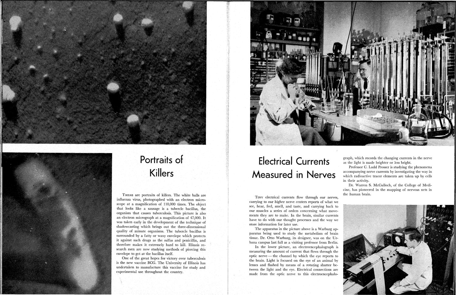
Portraits of Killers T H E S E are portraits of killers. T h e white balls are influenza virus, photographed with an electron microscope at a magnification of 110,000 times. T h e object that looks like a sausage is a tubercle bacillus, the organism that causes tuberculosis. This picture is also an electron micrograph at a magnification of 47,000. It was taken early in the development of the technique of shadowcasting which brings out the three-dimensional quality of minute organisms. T h e tubercle bacillus is surrounded by a fatty or waxy envelope which protects it against such drugs as the sulfas and penicillin, and therefore makes it extremely hard to kill. Illinois research men are now studying methods of piercing this envelope to get at the bacillus itself. One of the great hopes for victory over tuberculosis is the new vaccine BGG. T h e University of Illinois has undertaken to manufacture this vaccine for study and experimental use throughout the country. Electrical Currents Measured in Nerves T I N Y electrical currents flow through our nerves, carrying to our higher nerve centers reports of what we see, hear, feel, smell, and taste, and carrying back to our muscles a series of orders concerning what movements they are to make. In the brain, similar currents have to do with our thought processes and the way we store information for later use. T h e apparatus in the picture above is a Warburg apparatus being used to study the metabolism of brain tissue. Dr. Otto Warburg, its designer, was on the Urbana campus last fall as a visiting professor from Berlin. In the lower picture, an electroencephalograph is measuring the amount of current that flows through the optic nerve — the channel by which the eye reports to the brain. Light is focused on the eye of an animal by lenses and flashed by means of a rotating shutter between the light and the eye. Electrical connections are made from the optic nerve to this electroencephalo- graph, which records the changing currents in the nerve as the light is made brighter or less bright. Professor G. Ladd Prosser is studying the phenomena accompanying nerve currents by investigating the way in which radioactive tracer elements are taken u p by cells in their activity. Dr. Warren S. McGulloch, of the College of Medicine, has pioneered in the mapping of nervous nets in the human brain.
| |