| |
| |
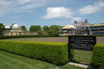
Caption: Board of Trustees Minutes - 1882
This is a reduced-resolution page image for fast online browsing.
EXTRACTED TEXT FROM PAGE:
128 DESCRIPTION OF PLATE I. [Fig. 1.—Micrococcus prodigiosus (Monasprodigiosa, Ehr.) Spherical bacteria of the red pigment, aggregated in pairs and in fours, the other pigment bacteria are not distinguishable with the microscope from this one. Fig. 2.—Micrococcus vaccinae. Spherical bacteria, from pock-lymph in a state of growth aggregated in short four to eight-jointed straight or bent chains, and forming also irregular cell-masses. Fig. 3.—Zooglcea-form of Micrococcus, pellicles or mucous strata characterized by granule-like closely set spherules. Fig. 4.—Eosary chain (Torulaform) of Micrococcus urese, from the urine. Fig. 5.—Kosary chain and yeast-like cell-masses from the white deposit of a solution of sugar of milk which had become sour. Fig. 6.—Sacharomyces glutinis (Cryptococcus glutinis, Fersen), a pullulating yeast which forms beautiful rose-colored patches on cooked potatoes. Fig. 7.—Sarcina spec, * from the blood of a healthy man, * * from the surface of a hen's egg grown over with Micrococcus luteus, forming yellow patches. Fig. 8.—Bacterium termo. free motile form. Fig. 9.—Zoogloea-form of Bacterium termo. Fig. 10.—Bacterium, pellicle, formed by rod-shaped bacteria arranged one against the other in a linear fashion, from the surface of sour beer. Fig. 11—Bacterium lineola, free motile form. Fig. 12.—Zoogloea-form of B. lineola. Fig. 13.—Motile filamentous Bacteria, with a spherical, or eliptical highly refringent "head," perhaps developed from gonidia. Fig, 14.—Bacillus subtilis, short cylinders, and longer, very flexible motile filaments, some of which are in process of division. Fig. 15.—Bacillus ulna, single segments and longer threads, some breaking up into segments. Fig. 16.—Yibrio rugula, single or in process of division. Fig. 17.—Yibrio serpens, longer or shorter threads, some dividing into bits, at * two threads entwined. Fig. 18.—"Swarm" of Y. serpens, the threads felted. Fig. 19.—Spirillum tenue, single and felted into "swarms." Fig. 20.—Spirillum undula. Fig. 21.—Spirillum volutans, * two spirals twisted around one another. Fig. 22.—Spirochaeta plicatilis. All the figures were drawn by Dr. Ferdinand Cohn with the immersion lens No. ix, of Hartnack Ocular in, representing a magnifying power of 650 diameters.]
| |